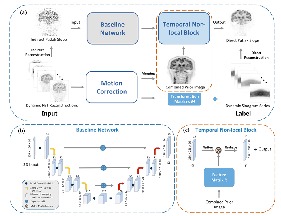
Rapid high-quality PET Patlak parametric image generation
Recently, deep neural networks have been widely and successfully applied in computer vision tasks and have attracted growing interest in medical imaging. One barrier for the application of deep neural networks to med- ical imaging is the need for large amounts of prior training pairs, which is not always feasible in clinical practice. This is especially true for medical image reconstruction problems, where raw data are needed. Inspired by the deep image prior framework, in this paper, we proposed a personal- ized network training method where no prior training pairs are needed, but only the patient’s own prior information. The network is updated during the iterative reconstruction process using the patient-specific prior information and measured data. We formulated the maximum-likelihood esti- mation as a constrained optimization problem and solved it using the alternating direction method of multipliers algorithm. Magnetic resonance imaging guided positron emission tomography reconstruction was employed as an example to demonstrate the effectiveness of the proposed framework. Quantification results based on simulation and real data show that the proposed reconstruction framework can outperform Gaussian post-smoothing and anatomically guided reconstructions using the kernel method or the neural-network penalty.
Related publications:
Rapid high-quality PET Patlak parametric image generation based on direct reconstruction and temporal nonlocal neural network, Neuroimage, 2021.
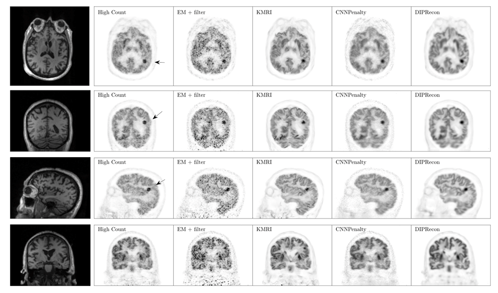
PET image reconstruction using deep image prior
Recently, deep neural networks have been widely and successfully applied in computer vision tasks and have attracted growing interest in medical imaging. One barrier for the application of deep neural networks to med- ical imaging is the need for large amounts of prior training pairs, which is not always feasible in clinical practice. This is especially true for medical image reconstruction problems, where raw data are needed. Inspired by the deep image prior framework, in this paper, we proposed a personal- ized network training method where no prior training pairs are needed, but only the patient’s own prior information. The network is updated during the iterative reconstruction process using the patient-specific prior information and measured data. We formulated the maximum-likelihood esti- mation as a constrained optimization problem and solved it using the alternating direction method of multipliers algorithm. Magnetic resonance imaging guided positron emission tomography reconstruction was employed as an example to demonstrate the effectiveness of the proposed framework. Quantification results based on simulation and real data show that the proposed reconstruction framework can outperform Gaussian post-smoothing and anatomically guided reconstructions using the kernel method or the neural-network penalty.
Related publications:
PET image reconstruction using deep image prior, IEEE Transactions on Medical Imaging, 2018.
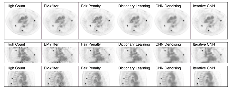
Iterative PET image reconstruction using convolutional neural network representation
PET image reconstruction is challenging due to the ill-poseness of the inverse problem and limited num- ber of detected photons. Recently, the deep neural networks have been widely and successfully used in computer vision tasks and attracted growing interests in medical imaging. In this paper, we trained a deep residual convolutional neural network to improve PET image quality by using the existing inter-patient information. An innovative feature of the proposed method is that we embed the neural net- work in the iterative reconstruction framework for image representation, rather than using it as a post-processing tool. We formulate the objective function as a constrained optimization problem and solve it using the alternating direction method of multipliers algorithm. Both simulation data and hybrid real data are used to evaluate the proposed method. Quantification results show that our proposed iterative neural network method can outperform the neural network denoising and conventional penalized maximum likelihood methods.
Related publications:
Iterative PET image reconstruction using convolutional neural network representation, IEEE Transactions on Medical Imaging, 2019.
Penalized PET Reconstruction using Deep Learning Prior and Local Linear Fitting
Motivated by the great potential of deep learning in medical imaging, we propose an iterative positron emission tomography reconstruction framework using a deep learning-based prior. We utilized the denoising convolutional neural network (DnCNN) method and trained the network using full-dose images as the ground truth and low dose images reconstructed from downsampled data by Poisson thinning as input. Since most published deep networks are trained at a predetermined noise level, the noise level disparity of training and testing data is a major problem for their applicability as a generalized prior. In particular, the noise level significantly changes in each iteration, which can potentially degrade the overall performance of iterative reconstruction. Due to insufficient existing studies, we conducted simulations and evaluated the degradation of performance at various noise conditions. Our findings indicated that DnCNN produces additional bias induced by the disparity of noise levels. To address this issue, we propose a local linear fitting function incorporated with the DnCNN prior to improve the image quality by preventing unwanted bias. We demonstrate that the resultant method is robust against noise level disparities despite the network being trained at a predetermined noise level. By means of bias and standard deviation studies via both simulations and clinical experiments, we show that the proposed method outperforms conventional methods based on total variation and non-local means penalties. We thereby confirm that the proposed method improves the reconstruction result both quantitatively and qualitatively.

Repository: https://github.com/kssigari/LLF
Related publications:
Depth-of-interaction Rebinning Strategy for
Ultrahigh Resolution PET
Small animal positron emission tomography (PET) imaging often requires high resolution (∼few hundred microns) to enable accurate quantitation in small structures such as animal brains. Recently, we have developed a prototype ultrahigh resolution depth-of-interaction (DOI) PET system that uses CdZnTe detectors with a detector pixel size of 350 μm and eight DOI layers with a 250 μm depth resolution. Due to the large number of line-of-response (LOR) combinations of DOIs, the systemmatrix for reconstruction is 64 times larger than that without DOI. While a high resolution virtual ring geometry can be employed to simplify the system matrix and create a sinogram, the LORs in such a sinogram tend to be sparse and irregular, leading to potential degradation of the reconstructed image quality. In this paper, we propose a novel high resolution sinogram rebinning method in which a uniform sub-sampling DOI strategy is employed. However, even with the high resolution rebinning strategy, the reconstructed image tends to be very noisy due to insufficient photon counts in many high resolution sinogram pixels. To reduce noise effects, we developed a penalized maximum likelihood reconstruction framework with the Poisson log-likelihood and a non-convex total variation penalty. Here, an ordered subsets separable quadratic surrogate and alternating direction method of multipliers are utilized to solve the optimization. To evaluate the performance of the proposed sub-sampling method and the penalized maximum likelihood reconstruction technique, we perform simulations and preliminary point source experiments. By comparing the reconstructed images and profiles based on sinograms without DOI, with rebinned DOI and with sub-sampled DOI, we demonstrate that the proposed method with sub-sampled DOIs can significantly improve the image quality with lower dose and yield a high resolution of <300 μm.

Related publications:
https://iopscience.iop.org/article/10.1088/1361-6560/aad58c/meta
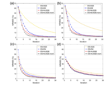
Time of Flight PET Reconstruction using Nonuniform Update for Regional Recovery Uniformity
Time of flight (TOF) PET reconstruction is well known to statistically improve the image quality compared to non-TOF PET. Although TOF PET can improve the overall signal to noise ratio (SNR) of the image compared to non-TOF PET, the SNR disparity between separate regions in the reconstructed image using TOF data becomes higher than that using non-TOF data. Using the conventional ordered subset expectation maximization (OS-EM) method, the SNR in the low activity regions becomes significantly lower than in the high activity regions due to the different photon statistics of TOF bins. A uniform recovery across different SNR regions is preferred if it can yield an overall good image quality within small number of iterations in practice. To allow more uniform recovery of regions, a spatially variant update is necessary for different SNR regions. This paper focuses on designing a spatially variant step size and proposes a TOF-PET reconstruction method that uses an non-uniform separable quadratic surrogates (NUSQS) algorithm, providing a straightforward control of spatially variant step size. To control the noise, a spatially invariant quadratic regularization is incorporated, which by itself does not theoretically affect the recovery uniformity. Nesterov’s momentum method with ordered subsets (OS) is also used to accelerate the reconstruction speed. To evaluate the proposed method, an XCAT simulation phantom and clinical data from a pancreas cancer patient with full (ground truth) and 6x downsampled counts were used, where a Poisson thinning process was employed for downsampling. We selected tumor and cold regions of interest (ROIs) and compared the proposed method with the TOF-based conventional OS-EM and OS-SQS algorithms with an early stopping criterion. In computer simulation, without regularization, hot regions of OS-EM and OS-NUSQS converged similarly, but cold region of OS-EM was noisier than OS-NUSQS after 24 iterations. With regularization, although the overall speeds of OS-EM and OS-NUSQS were similar, recovery ratios of hot and cold regions reconstructed by OS-NUSQS were more uniform compared to those of the conventional OS-SQS and OS-EM. The OS-NUSQS with Nesterov’s momentum converged faster than others while preserving the uniform recovery. In the clinical example, we demonstrated that the OS-NUSQS with Nesterov’s momentum provides more uniform recovery ratios of hot and cold ROIs compared to OS-SQS and OS-EM. Although the cost function of all methods are equivalent, the proposed method has higher structural similarity (SSIM) values of hot and cold regions compared to other methods after 24 iterations. Furthermore, our computing time using graphics processing unit was 80x shorter than the time using quad-core CPUs. This paper proposes a TOF PET reconstruction method using OS-NUSQS with Nesterov’s momentum for uniform recovery of different SNR regions. In particular, the spatially non-uniform step size in the proposed method provides uniform recovery ratios of different SNR regions, and Nesterov’s momentum further accelerates overall convergence while preserving uniform recovery. The computer simulation and clinical example demonstrate that the proposed method converges uniformly across ROIs. In addition, tumor contrast and SSIM of the proposed method were higher than those of conventional OS-EM and OS-SQS in early iterations.
Related publications:
https://aapm.onlinelibrary.wiley.com/doi/abs/10.1002/mp.13321
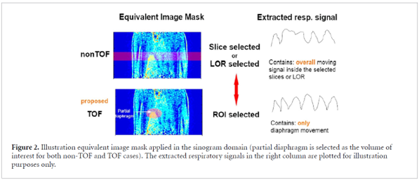
Data-driven Respiratory Gating based on Localized Diaphragm Sensing in TOF PET
It is important to measure the respiratory cycle in positron emission tomography (PET) to enhance the contrast of the tumor as well as the accuracy of its localization in organs such as the lung and liver. Several types of data-driven respiratory gating methods, such as center of mass and principal component analysis, have been developed to directly measure the breathing cycle from PET images and listmode data. However, the breathing cycle is still hard to detect in low signal-to-noise ratio (SNR) data, particularly in low dose PET/CT scans. To address this issue, a time-of-flight (TOF) PET is currently utilized for the data-driven respiratory gating because of its higher SNR and better localization of the region of interest. To further improve the accuracy of respiratory gating with TOF information, we propose an accurate data-driven respiratory gating method, which retrospectively derives the respiratory signal using a localized sensing method based on a diaphragm mask in TOF PET data. To assess the accuracy of the proposed method, the performance is evaluated with three patient datasets, and a pressure-belt signal as the ground truth is compared. In our experiments, we validate that the respiratory signal using the proposed data-driven gating method is well matched to the pressure-belt respiratory signal with less than 5% peak time errors and over 80% trace correlations. Based on gated signals, the respiratory-gated image of the proposed method provides more clear edges of organs compared to images using conventional non-TOF methods. Therefore, we demonstrate that the proposed method can achieve improvements for the accuracy of gating signals and image quality.
Related publications:
https://iopscience.iop.org/article/10.1088/1361-6560/ab9660/meta